In this blog, we explore the detection of multiple proteins and discuss the fluorescence multiplexing detection method.
Detecting multiple protein molecules on a Western blot can save time, cost, and resources in the laboratory. In this blog post, we explore the detection of multiple protein molecules for protein quantitation in Western blotting and discuss what the fluorescence multiplexing detection method has to offer.
Why detect multiple protein molecules in Western blotting with fluorescent?
If you’re an immunoblotting expert, detecting more than one protein molecule on a single Western blot can be an experimental necessity. Stripping Western blots and reprobing the blot is common practice, but this process has its drawbacks.
When multiplexing with fluorescent probes, you can detect multiple protein molecules simultaneously, even those with similar molecular weights.
Difference between Fluorescent vs chemiluminescent probes
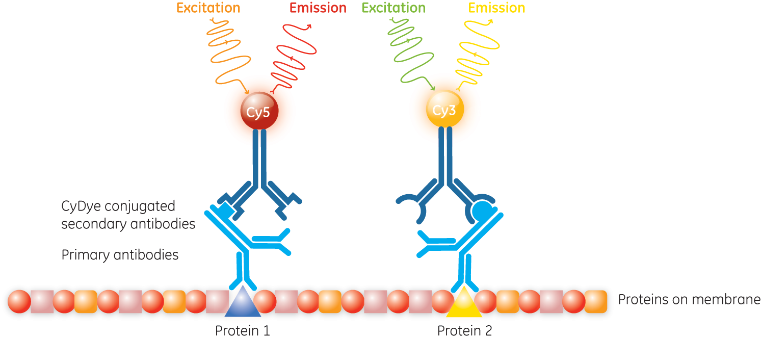
Using multiplexed fluorescence in your Western blots uses different fluorescently conjugated antibodies (for example, Cy3 and Cy5 fluorescent dyes) to target and detect protein molecules. Multiplexing is an alternative to membrane stripping and reprobing, in that you probe for your target protein molecule in much the same way, but can add multiple antibodies at the same time (Fig 1).
On the other hand, stripping chemiluminescent antibodies and reprobing a single membrane is not a true multiplex. Stripping in Western blotting also carries a risk of protein molecule loss, which undermines confidence in normalization or quantitation of the protein molecules.
Western blot membrane stripping can also involve harsh conditions such as high pH and temperatures. When your target protein molecule is in low abundance, losing some of it might make it undetectable. For more information on measuring the weight of your Western blot molecules, read our blog on Molecular markers for polyacrylamide gel electrophoresis here.
If your proteins are different molecule sizes, you might be able to cut your membrane in two and probe each piece with a different antibody, but this won’t help if you’re looking for similarly sized protein molecules.
Fig 1. The principle of Amersham ECL Plex for fluorescent Western blot. Primary antibodies against two proteins are recognized by species-specific secondary antibodies conjugated to different Amersham CyDye fluorescent dyes.
The advantages of fluorescence Western Blots
If you are detecting fluorescent signals directly from fluorophore-labeled secondary antibodies, you won’t need enzyme or substrate reagents. This signal is also stable and doesn’t decay much, which means it has the potential to be detectable for months or even years – saving valuable resources!
Fluorescence Western blots is a sensitive protein molecule detection method where the signal is proportional to protein quantity. This property gives fluorescence a broad linear dynamic range, allowing you to study high- and low-abundance proteins at the same time.
These properties make fluorescence detection in Western blots an excellent choice for quantitative, multiple protein detection and analysis.
Multifluorescence examples
The two following examples demonstrate the power of multiplexed fluorescence in Western blotting for the detection of multiple protein molecules.
Detecting proteins with similar molecular weights
This first example uses Amersham ECL Plex to visualize protein molecules that differ in size only by a post-translational modification. It is possible to use a fluorescence multiplex to monitor the expression of unmodified and modified protein forms simultaneously by detecting two epitopes on a single protein.
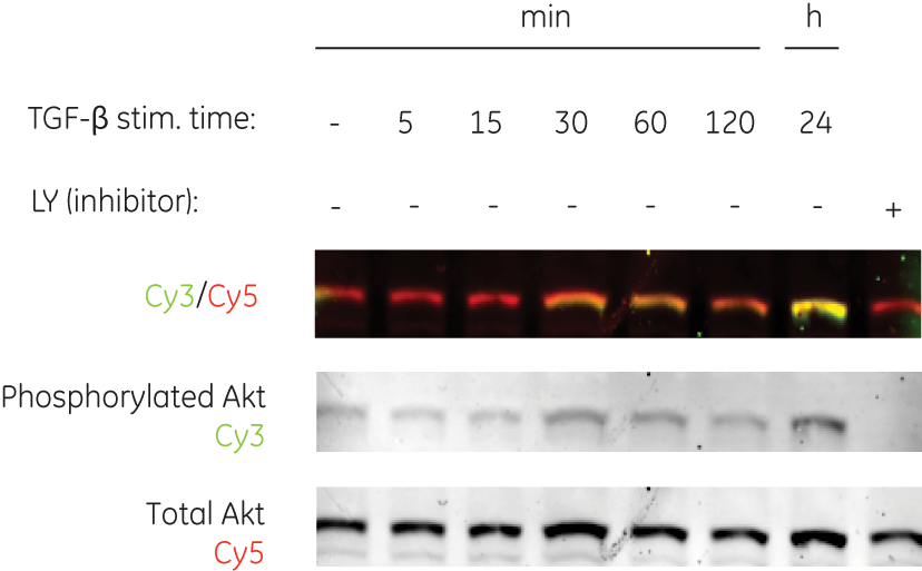
Multiplexing is particularly handy for looking at protein phosphorylation, as illustrated in this example (Fig 2). Although this modification causes only minimal changes in molecular weight of the protein molecules, using two fluorescent probes allows you to discriminate between the two forms – saving countless work hours.
Fig 2. Multiple protein molecule detection of two forms of human Akt protein (phosphorylated and total) using Amersham ECL Plex. After blocking and probing with anti-Akt and anti-phosphoAkt primary antibodies, the membrane was probed with two Amersham ECL Plex secondary antibodies: an anti-mouse Cy5 and anti-rabbit Cy3. Despite the minimal change in molecular weight as a result of phosphorylation, the duplex capability of Amersham ECL Plex enables a clear distinction between the two forms of the protein. Note the complete absence of signal in the Cy3 channel for the sample treated with kinase inhibitor, LY. Data courtesy of Marene Landstrom, Ludwig Institute for Cancer Research, Uppsala, Sweden.
Triplexed protein detection – Comparison of two protein molecules with total protein normalization
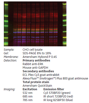
Here we take multiplexed detection up a notch to compare expression of two proteins normalized to a housekeeping protein molecule. This brings the number of proteins detected on a single membrane to three, each with a distinct fluorophore (Fig 3). The fluorophores emit signals at discrete wavelengths, enabling simultaneous detection and analysis.
Fig 3. Triplex protein detection with total protein normalization. Different amounts of CHO cell lysate were loaded on an SDS-PAGE gel for Western Blot detection. ERK and GAPDH were detected using the 532 nm and 785 nm lasers respectively.
Considerations for multifluorescence
Fluorescence detection Western blot expands the technique’s protein molecule identification and quantitation capabilities.
In this post, we’ve talked about multiplexing to detect up to three targets. Of course, you could continue to increase the number of probes to four or more, but you must carefully consider the experimental design as each primary antibody would need to be raised in a different, or at least distinguishable, host.
Additionally, fluorescence imaging depends on having an imaging device with appropriate filters built in. Laser scanners, such as Typhoon, come with several options for filters, which are expandable as needed.
So, the next time you run an experiment involving a Western blot, and find yourself targeting and detecting multiple protein molecules, consider multifluorescence. This method enables you to detect multiple proteins simultaneously without the risk of protein loss from stripping and reprobing your Western blot experiment.
For information on how to prepare Western blot buffers, check out our Western Blotting Principles and Methods Handbook.


