Comparison of transfer methods in Western blotting
After gel electrophoresis, Western blotting starts with the transfer of proteins from the gel to membrane. Electrotransfer is a common technique for step and takes advantage of the same electromobility principles as gel electrophoresis.
Wet and semidry transfer are the two most common electrotransfer methods and provide greater speed and efficiency than alternatives based on diffusion, capillary action, and vacuum.
What is wet transfer?
Wet transfer is generally suitable for all proteins and uses a cassette that sandwiches foam pads, filter paper, gel, and a membrane together. The cassette is submerged in transfer buffer and orientated with the membrane on the anode side (Fig 1), to ensure proteins move in the correct direction.
During electrophoresis, negatively charged proteins migrate from the gel towards the anode, contacting and binding to the membrane. Transfer speed ranges from 1 h to overnight, usually at a constant voltage to maintain field strength and maximize efficiency of transfer.
Long transfer times at a constant voltage lead to unwanted heat, decreasing resistance, and increasing current. Adequate cooling measures to prevent buffer breakdown are often required, including chilled buffers, ice packs, and performing transfers in a cold room at 4°C. Some wet transfer systems use integrated cooling.
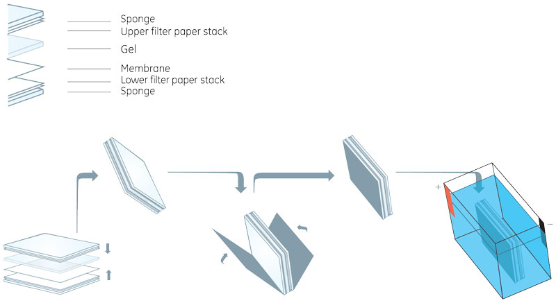
Fig 1. The wet transfer sandwich is assembled (top), placed in a cassette, and submerged in a transfer tank filled with buffer with membrane closest to the anode (bottom).
What is semidry transfer?
Semidry transfer follows the same principles as wet transfer but is faster and uses smaller volumes of buffer.
The gel and membrane are sandwiched between sheets of filter paper soaked in transfer buffer, with electrode plates on either side (Fig 2). As in wet transfer, proteins move out of the gel toward the anode when an electric field is applied, binding to the membrane.
Unlike wet transfer, semidry transfer uses minimal buffer and requires minutes, compared to hours. The electrode plates absorb heat during the short run time, necessitating fewer cooling measures. However, semidry transfer is less efficient overall, noticeably affecting the transfer of high molecular weight proteins.
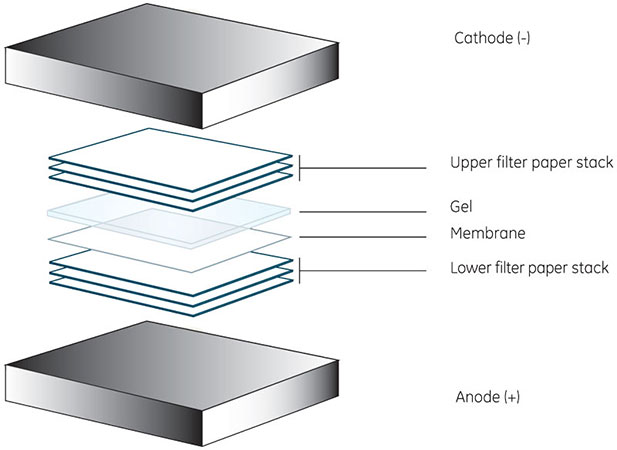
Fig 2. Construction of a gel-membrane sandwich in a semidry transfer setup is similar to wet transfer. All components are soaked in transfer buffer and placed in direct contact with the electrodes, compressing the sandwich.
Following either wet or semidry transfer, the membrane can be stained or immunoblotted, as needed.
What are the advantages/disadvantages of these methods?
Table 1 summarizes the main differences between wet and semidry transfer.
Table 1. Comparison of wet and semidry transfer of proteins
| Advantages | Disadvantages | |
| Wet transfer | Efficient transfer for small and large proteins | Requires adequate cooling measures |
| Clear, sharp bands on membrane | Large volumes of buffer | |
| Particularly suited for transfer of large proteins | Running times from 1 h to overnight | |
| Semidry transfer | Short run times (<1 h) | Lower overall transfer efficiency than wet transfer |
| Requires small buffer volumes | Unsuitable for large proteins | |
| Efficient transfer of small proteins (Mr 20 000), particularly with polyvinyl difluoride (PVDF) membranes | Tendency to produce indistinct bands on membrane |
Which method should I choose?
Protein size is a significant factor in the choice of transfer method. The inherently slower migration of large proteins can lead to poor transfer efficiency in the semidry method. However, the speed of semidry transfer makes it convenient for experiments that require a quick check for the presence or absence of a protein.
In contrast, for proteins larger than relative molecular mass (Mr) 80 000 or those requiring quantitative analysis, the extended transfer times of wet transfer are a more suitable option. Figure 3 highlights the difference in efficiency between wet and semidry transfer of human transferrin (Mr 80 000).
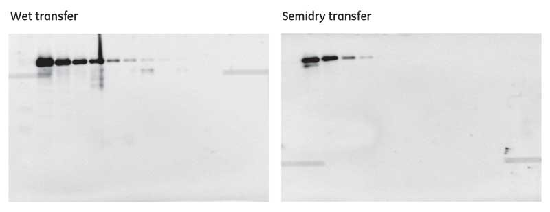
Fig 3. A two-fold dilution series of transferrin (bands from left to right), starting at 5 µg, transferred by wet (left) and semidry (right) methods to a PVDF membrane. Semidry transfer Semidry transfer is less efficient for this particular protein than wet transfer; less protein throughout the dilution series is transferred, leading to reduced sensitivity.
For more information, download the Western blotting handbook.