Scientists often use a UV detector to measure the total protein content of eluting peaks in chromatography. The amount of light absorbed is directly proportional to the concentration of proteins in the solution and follows Beer-Lambert’s law, A=ε*l*c, where A is the absorbance, ε is the molar extinction coefficient or absorptivity of the attenuating species, l is the optical path length in cm, and c is the concentration of the attenuating species. The UV flow cell pathlength defines the volume of the sample exposed to the analyzer’s light beam — this means that adjusting the length of the flow cell determines how much of the sample is being measured. Selecting the right flow cell pathlength is critical to ensure that absorbance values are within the (linear) range of the detector. Scientists choose the flow cell pathlength based on absorbance of the lowest and highest expected concentration so that they can make valid calculations for the protein concentrations.
Read on to learn how you can determine the right flow cell pathlength.
UV absorption of proteins at 280 nm is mainly due to the presence and number of aromatic residues from amino acids tryptophan (Trp) and tyrosine (Tyr). Each individual protein has its own distinct absorption spectra and absorption coefficient. To make sure the sample concentration is within the dynamic range of the detector (typically 0 – 2000 mAU), we must choose the right flow cell pathlength.
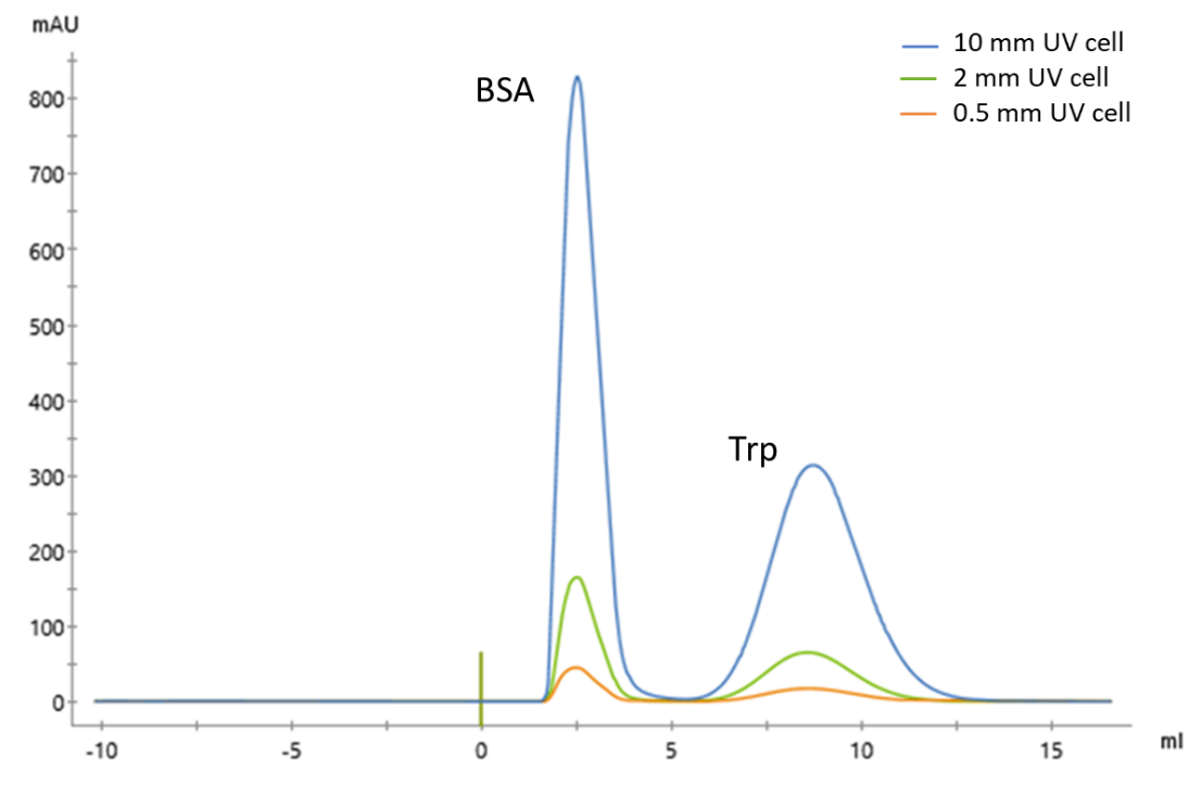
Figure 1: UV data from chromatographic runs of bovine serum albumin (BSA) (1.5 mg/mL) and Trp (0.04 mg/mL) performed on a ÄKTA pure™ instrument equipped with flow cells with 0.5 mm, 2 mm and 10 mm pathlength.
What if the eluting protein concentration is low and the UV peak is difficult to detect?
If the peak detection and subsequent fractionation is difficult due to low peak intensity, it means the protein concentration passing through the flow cell is below the detection range of the UV module. To increase the number of absorbing proteins, we can consider increasing the UV flow cell pathlength. Increasing the UV flow cell pathlength will increase the sample volume of the eluting peak you’re analyzing, and bring the UV signal within the detectable range of the UV detector. This supports better concentration estimates and eventually, more accurate fractionation of the peak.
For example: If you’re using a 2 mm UV flow cell, replace it with a 10 mm UV flow cell. Figure 1 shows the difference in the UV response for the same molecules with different UV flow cell pathlengths. UV intensity amplifies about five-fold on a 10 mm UV flow cell compared to the standard 2 mm UV flow cell.
What if protein concentration is too high and difficult to quantitate?
If the protein concentration is very high and your data doesn’t make sense, it means the detected concentration is beyond the linear detection range of the UV module. To obtain UV data within the linear dynamic range of the detector, you need to bring down the UV flow cell pathlength. Decreasing the UV flow cell pathlength will decrease the amount of you’re analyzing and bring the UV signal within the dynamic range of the detector, helping you more accurately detect and quantitate the peak.
For example, if you’re using a 2 mm UV flow cell, replace it with a 0.5 mm UV flow cell. Figure 1 shows the difference in the UV response for the same molecules with different UV flow cell pathlengths. The UV intensity reduces about four-fold on a 0.5 mm UV flow cell compared to the standard 2 mm UV flow cell.
How can we detect proteins at high and low concentrations at the same time?
You can use more than one UV module on your chromatography system to extend the detection range. Using two UV flow cells with two different flow cell pathlengths will extend the detection range and help you better monitor eluting proteins of different concentrations (Figure 2).
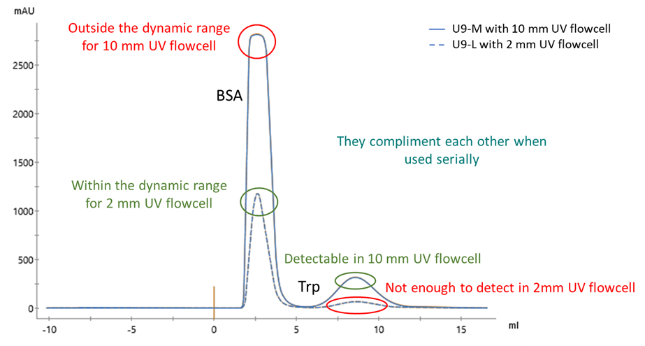
Figure 2: UV curve of BSA (12 mg/mL) and Trp (0.032 mg/mL) on ӒKTA pure™ instrument with U9-M UV module fitted with 10 mm UV flow cell and U9-L UV module fitted with 2 mm UV flow cell.
In conclusion, your selected UV flow cell path length depends on the protein concentration in your sample and its eluting peak. If your protein concentration is very low and below the detection range of the UV module, it might be enough to select a UV cell with a longer pathlength. If the protein concentration is very high — beyond the linear dynamic range of the UV module — then you either need to select a shorter pathlength or dilute your sample. You can also use more than one UV flow cell with different pathlengths to extend your chromatography system’s detection range and get more accurate detection of peaks and data analysis over a wide concentration range.