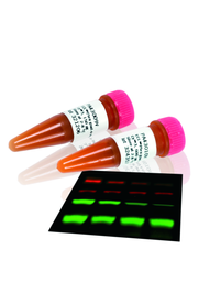
ECL Plex Goat-α-Rabbit IgG-Cy5, for 1000 cm² membrane area
Amersham ECL Plex Cy3 and Cy5 labeled antibodies are part of an optimized system for quantitative analysis using fluorescent Western blotting.
FAQ
I have used ECL Plex and I have always a lot of background in the Cy3 channel. So ECL Plex Cy3 does not work so well for me. What can be the problem?
For lowest background, we recommend to scan the membranes dry. Also the scan setting is important. If you use a Typhoon 8600, the Cy3 filter setting will automatically be 555 BP 20, which is suboptimal and give high background and weak signals. Make sure to use the 580 BP 30 Cy3 TAMRA filter setting for optimal Cy3 detection.
I do not want to mix two primary antibodies in the same tube to perform a multiplex ECL Plex experiment, how can I overcome this?
You can perform the probing one after the other, and in this way avoid mixing the antibody solutions.
If I label the primary antibody is there not a risk that the binding site is affected?
Yes, the binding affinity of the antibody can be affected if the Dye is conjugated to or close to the binding domain of the antibody. It is important to follow the protocol for antibody labeling so it will be an optimal number of dye molecules incorporated per antibody.
Primary antibody detection should be less sensitive compared to primary and secondary antibody detection? Will the signal be as strong when using only labled primary antibodies?
Yes, the signal will be amplified with each probing layer, so if you only have one layer, the signal may be a little lower. So use this detection for targets of medium to high abundance. Avoid it for low abundant targets.
How do I know that the membrane is low fluorescent? And, can I use the same membrane for 2D western?
How do I know that the membrane is low fluorescent? And, can I use the same membrane for 2D Western Blotting?
We have 2 low fluorescent membranes that we recommend for ECL Plex. One NC, Hybond ECL and one PVDF alternative called Hybond LFP. You can try other membranes, but then you have to test them first. If they give elevated background, there is risk that you loose the weak signals. And yes, you can also use them for 2D Western Blotting.
I have had problems when I scan my ECL Plex blots. I sometimes see areas that are much lighter and have weaker signals. Why is that?
Most likely, this is caused by partial drying of the membrane. The areas that have dried give very different intensities compared to the ones that are still wet. The membrane can start to dry on the scan bed. We recommend to always dry the membranes before scanning for best results (lower background and better signal to noise ratio).
When are stable signals important I only need to detect my signals one time anyway?
Stable signals improve reproducibility and the possibility for you to analyze many blots in parallel. If you for example have 5 blots, it is very hard to react and detect with exactly identical timings. If the signal vary within minutes depending on were in the enzyme (HPR) reaction time curve you are it is impossible to get accurate data. Also, if you want to add blots to your experiment or want to compared with previous data, for example blots from the previous week, it is very helpful with stable signals. If you work with ECL Plus chemifluorescence of ECL Plex fluorescence you will get very stable signals you have the possibility of processing many blots and compare your results between different experiments.
Can I use fluorescent Western Blotting with ECL plex if I have human primary antibodies?
Yes, it is possible if you label the primary antibody yourself using an antibody labeling kit from Cytiva.
Can you strip and re-probe ECL Plex probed blots?
Yes, we have detailed information in the ECL Plex protocol of a compatible stripping solution containing ß-mercaptoethanol. Usually, you do not need to strip and re-probe ECL Plex blots, since you can do multiplexing directly.
How can I optimize my antibody dilutions in an easy way?
Traditionally the dot blot method is used for determining the optimum dilution of antibodies. A more reliable method that mimics a true experiment is to prepare a Western blot and divide it into strips/sections. This takes longer time than dot blots, but more parameters, such as specific signal intensity, background and amount of unspecific detection can be monitored.
By using this method you will be able to chose:
- Optimal dilutions of primary and secondary antibodies
- Optimal sample load
- Optimal blocking agent
- Best species (e.g. rabbit or mouse) of primary antibody
- Best quality of primary antibody (choice of best supplier)
Is there a problem with incubation of 2 antibodies simultaneously? Is there a risk that the antibodies compete/interfere with one another?
There is a risk if you have epitopes really close together. In most cases this is not a problem. If you know that your epitopes are really close, you can probe 2 blots separately and multiplex with a housekeeping protein instead and then combine the data and get reliable quantitative results.
Why is the detection limit lower (4.9 pg compared to 9.8 pg) using film on one slide compared to a previous slide?
It can be explained by experimental variation caused by transfer, dilution series, primary antibody affinity (different lots) etc.
What is the definition of limit of detection?
When a protein band has a signal to noise ratio above 3, i.e. the signal is 3 times larger than the variation in background.
Do you have more species of your ECL Plex antibodies?
There is a limitation of detecting two different species, rabbit and mouse with ECL Plex. But you can always label the primary antibodies from other species by using our antibody labeling kits. If you can label a third species of primary antibody with one of our antibody labelling kits in addition to rabbit and mouse primary antibodies. In this way use all 3 CyDyes also for ECL Plex and perform a triplexed detection.
Stripping and re-probing of membranes is time consuming and the results are uncertain if we have lost proteins unevenly across the blot during the stripping procedure. What can I do to avoid this if I have 2 targets of the same size?
If you work with chemiluminescence, you can probe 2 blots in parallel with the same samples loaded. One blot is probed for 1 target + housekeeping protein, the other blot for the second target + housekeeping protein. The normalized signals for target 1 and 2 can be compared if the blots were reacted and detected simultaneously.
For ECL Plex, you can perform multiplex detection of the targets simultaneously and detect the 2 targets separately in different channels. For normalization to house-keeping protein you can perform triplex detection.
Can I use the same primary antibody dilutions for ECL plex as I use for ECL plus?
Usually, optimal dilution of primary antibodies is the same or somewhat lower for ECL Plex compared to ECL Plus. When optimizing, I would try the same as for ECL Plus and 2 lower dilutions for ECL Plex. Usually, as with other fluorescent detection systems, ECL Plex requires higher concentrations of primary antibodies.
Troubleshooting
Find solutions to product related issues. For unlisted issues please contact local Cytiva service representation.
Issues related to detection of low abundant proteins
| Possible cause | Suggested remedy |
|---|---|
- |
Use ECL Plex Cy5 conjugated secondary antibody for the protein you expect to have the lowest concentration in your sample. The ECL Plex Cy5 secondary antibody is slightly more sensitive than the ECL Plex Cy3 secondary antibody and the ECL Plex Cy2 secondary antibody. |
- |
When detecting low abundant proteins, the signal can for a few target proteins become stronger if the species of primary antibody is switched in addition to careful optimization of antibody dilution. Sometimes the signal for the protein of interest can become stronger using an anti-mouse instead of an anti-rabbit primary antibody and vice versa. |
- |
For improved detection of weak signals using the Hybond-LFP membrane, skip the blocking step and instead dry the membrane directly after transfer. Dry the membrane for at least 2 hours at ambient temperature. |
- |
If you are detecting low abundant proteins, use a smaller amount of ECL Plex Rainbow Markers (1.5 μl) or leave an empty lane between the markers and the sample. The markers can otherwise appear too strong and even disturb the analysis of the sample lane. |
- |
TBS (Tris buffered saline) and TBS-T is recommended for use with antibodies against phosphorylated proteins. The phosphorate in PBS buffer can interfere and reduce the antibody binding. |