Are you having trouble using ECL products for Western blotting (immunoblotting)? See our tips and tricks for Western blot detection.
Probing and detection is the final step in the long process of Western blotting, and unfortunately, there are still things that can go wrong at this stage.
Here are some of the snags we sometimes hear about from researchers using enhanced chemiluminescence (ECL) products, and some tips to help fix them.
Weak or no signal
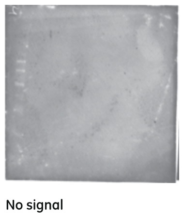
There are three main reasons for not seeing visible bands on your membrane: poor protein transfer, problems relating to the primary or secondary antibody, or problems with the detection reagent.
To confirm protein transfer, stain your membrane with a total protein stain such as Ponceau S. If the transfer was successful check that the antibodies and ECL reagents were stored correctly and used at the right concentrations. Also, check the storage conditions of the blots after transfer.
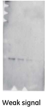
When the bands are visible but appear weak, the issue could be low protein loading in the gel, poor transfer, too little or inactive antibody, or short exposure times (Fig 1).
Remember that an antibody raised against a native protein might not bind the denatured form. Further, make sure the secondary antibody is directed against the species in which the primary antibody was raised. Verify the concentration of your antibodies using, for example dot-blotting. Strip and reprobe the membrane if necessary.
Fig 1. No signal (A) and weak signal (B) in Western blotting.
Excessive, diffuse signals
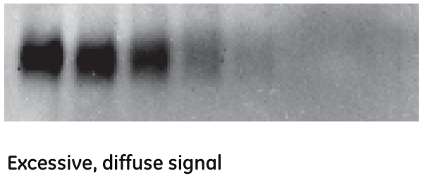
Excessive amounts of protein or high concentrations of antibodies, but also excessive exposure times, can lead to saturated signals. These signals are no longer proportional to protein concentration and may therefore not be used for quantitation of molecular weight. The high, diffuse signal intensity also makes it difficult to resolve protein bands that are close together. Reduce the concentration of the antibodies, or simply load less protein onto your gel to avoid diffuse signals.
Fig 2. Excessive, diffuse signal in ECL detection.
Negative bands on the film
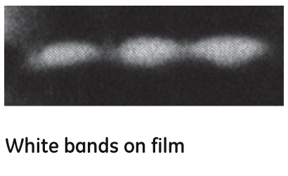
Occasionally, when imaging a membrane with chemiluminescence, white (negative) bands can appear alongside the normal black bands (Fig 3). These are the result of substrate depletion and it becomes difficult to quantitate the band in question.
Substrate depletion occurs when too much of the enzyme horseradish peroxidase (HRP) is present in a band, quickly consuming most of the substrate. Reducing either the target protein or the antibody concentration should help prevent white bands.
Fig 3. White bands on film.
Spotted (speckled) background
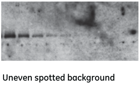
Spotted backgrounds (Fig 4) are a problem that can be caused by problems with the blocking agent, antibody, or contaminated equipment.
Ensure that the blocking agent is dissolved in the buffer by gentle warming and mixing to avoid aggregates. Aggregates in the HRP-conjugated antibody can also cause speckling; filter through a 0.2 µm filter.
Always keep surfaces in contact with the membrane clean. Wiping the surface of the scanner with 70% ethanol followed by deionized water is good practice.
The golden rules are to handle the membrane with care and with clean forceps.
Fig 4. Uneven, spotted background.
High background
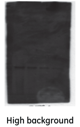
Once again, there are several reasons why the background can be higher than you want it to be (Fig 5). This might result from insufficient blocking time or low concentration of blocking agent. Simply increase the blocking time and/or concentration of blocking agent.
Concentration of antibodies and concentration of detergent in the washing buffer are a couple of typical causes. Try reducing the concentration of antibodies or reducing the concentration of detergent. Also, ensure that that the blocking and washing buffers are freshly prepared.
Contamination is another important point. Make sure you are using clean transfer and incubation buffers. Also, try not to cut corners on antibody washes, as insufficient washing is a frequent source of background staining in antibody-based detection
Fig 5. High background.
These are some of our tips for troubleshooting Western blots. If you are still experiencing problems, find more hints and tips in the Western blotting principles and methods handbook.
