Blood separation is a key part of many diagnostic assays. Removing red and white blood cells reduces the red coloration and eliminates interference from intracellular material, such as DNA.
Diagnostic assays that measure an analyte in blood plasma often use filters for separation, also known as blood separation membranes, most commonly taking advantage of the properties of depth filters.
How depth filters separate blood particles
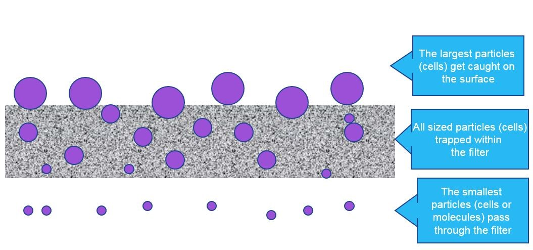
Depth filters work by trapping particles both inside and on the surface of the filter (Fig 1A). The largest particles remain on the surface, whereas smaller particles become trapped within the fiber mesh of the depth filter. This gradual filtration effect reduces clogging of the filter material, enabling rapid filtration and use of high particle loads.
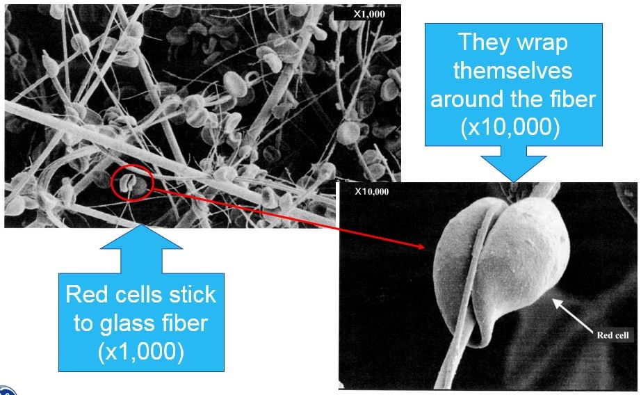
Filters with a glass fiber matrix are well suited for use in blood separation assays. They are available in a wide variety of thicknesses, enabling developers to accommodate a range of blood volumes, depending on the assay needs. Glass fiber has the additional benefit that red blood cells bind to the surface of the glass, wrapping themselves around the fiber (Fig 1B).
A
B
Fig 1. Separation of particles by depth filter. (A) The largest particles remaining on the surface of the filter; (B) Red blood cells binding to glass fibers.
Properties of a Blood Separator
Characteristics of a depth filter essential to blood separation include its filter area and direction of flow. The required filter area depends largely on the expected sample volume. Small volume samples need a small filter area to minimize the hold-up volume, whereas large volume samples need a large surface area to prevent clogging.
There are three options for the direction of flow: vertical, lateral, and composite.
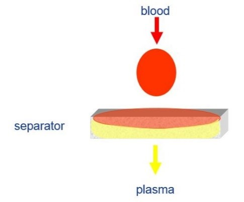
Vertical flow filtration (Fig 2A) involves placing a blood sample on top of a filter, which is then separated as it passes through the filter matrix. Vertical flow is suitable for high-volume applications requiring fast separation.
Key drawbacks of this method include low filtration efficiency and low serum recovery, which could produce false results in a diagnostic assay.
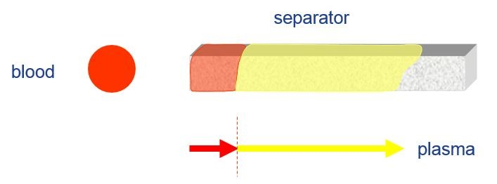
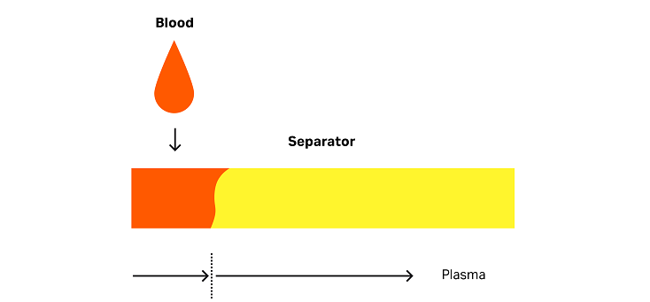
Lateral flow filtration (Fig 2B) is the principle behind so-called ‘dipstick’ assays. In this type of diagnostic assay, the operator immerses part of the filter matrix in a blood sample. The matrix then separates the cells from the plasma as the sample travels through the material.
This approach is well suited for diagnostic assays involving small sample volumes. Typically, these assays have a high efficiency, and serum recovery is generally over 85%. These filters, however, clog easily with sample volumes greater than 100 µL and the filtration process is often slower than for vertical flow.
Composite flow consists of a vertical blood separation phase followed by lateral separation (Fig 2C). In a composite setup the filter matrix sits inside a cassette housing with an opening at the top sample application. After application, the sample travels vertically into the matrix and then laterally through the matrix.
This approach combines some of the benefits of vertical and lateral flow. Composite assays have high filtration efficiency and sample recovery, but are also able to cope with larger sample volumes than a typical dipstick assay. This is the typical blood separation procedure in a cassette-based lateral flow diagnostic test.
Vertical flow
Lateral flow
Composite flow
Fig 2. Three blood separation methods. (A) Comparison of vertical flow; (B) lateral flow; and (C) composite flow.
Read more about challenges that can prevent efficient blood separation in diagnostic assays and potential solutions or download our brochure to learn more about building a diagnostic assay.
Unsure of which depth filtration material to use?
Cytiva's team of experts are trained in troubleshooting filtration. They can advise on which types of filters are suitable in your assay, and help identify the cause of and solution to any issues faced in diagnostic assay development. Contact your local Cytiva representative to discuss your needs.
Read Part 1 - Blood Separation Challenges (and How to Solve Them!)
For Samples or to contact a Specialist
Related Documents:
- Brochure: Build a Smarter Diagnostic Assay
- Infographic: Diving into Diagnostics
Tools:
- Whatman Filter Selector
- Typical Properties of Filter Papers and Glass Fiber Filters