Sanitization methods using 20 mM peracetic acid (PAA) as sanitization agent were evaluated in the packing and repacking of a protein A resin in AxiChrom 300 column. The sanitization agent PAA is an oxidizing agent that effectively removes both vegetative and spore-forming bacteria.The goal of the study was to ensure sanitization in the no-flow zone between bed supports and column tube of the AxiChrom column.
The column was packed with prechallenged MabSelect SuRe base matrix using Pseudomonas aeruginosa as challenging organism. Highly efficient sanitization in place during the packing procedure was observed by modification of the sanitization method to ensure permeation of column no-flow zones with the sanitization agent. The level of P. aeruginosa was reduced from 107 colony forming units (CFU)/mL of the challenging organism to levels of no detection throughout the whole packed bed including resin in the space between bed supports and tube.
The sanitization method described was developed in collaboration with Roche.
- Introduction
- Microbial challenge test
- Materials and sanitization methods
- Preparation of system and pre-sanitization of column
- Packing and sanitization of AxiChrom 300 column
- Sanitization of challenged column
- Liquid sample collection
- Microbial sampling
- Results from the sanitization procedure
- Bed performance after sanitization
- Conclusions
- Trademark information
Introduction
Minimizing bioburden through use of sodium hydroxide (NaOH) and avoiding contamination are highly prioritized by biopharmaceutical manufacturers to avoid high costs of resolving these issues and ensure no delays in the downstream process. For chromatography resins with protein ligands that are sensitive to high pH conditions, lower concentrations of NaOH or alternative cleaning and sanitization solutions must be used. Experience has resulted in 1 M NaOH becoming the backbone of most sanitization procedures, due to efficacy and low cost as well as ease of detection, removal, and disposal.
The use of 1 M NaOH for clearance/sanitization of spores is however not as effective as it is for vegetative contaminants. As a result, a collaboration with Roche was initiated with the goal to devise an alternative sanitization method during packing or remediation of a packed bed of MabSelect SuRe resin in AxiChrom columns using peracetic acid (PAA). The sanitization agent PAA is an oxidizing agent that effectively removes both vegetative and spore-forming bacteria. PAA is compatible with MabSelect SuRe resin and most hardware bioprocess equipment. Previous investigations have shown that treatment of MabSelect SuRe resin with 20 mM PAA for 30 min or 30 mM PAA for 15 min can be used without significantly affecting purification performance of the resin. In a long-term study, MabSelect SuRe resin was subjected to 20 mM PAA in a total of 28 treatments with a total contact time of 14 h over 108 protein A process cycles, without significant effect on performance.
Microbial challenge test
The principle of microbial challenge testing is to introduce high concentrations of microorganisms into the equipment (e.g., the column) to be investigated. The equipment is then treated with the sanitizing/cleaning agent. Following treatment, samples are taken at predetermined sites, and the numbers of viable cells remaining are measured.
The goal of the study was to describe a best practice in column handling and sanitization and the best practice in terms of sanitization of the protein A resin throughout the entire packed bed, including trapped resin in the no-flow zones between bed supports and tube. A novel aspect of this study was that contamination was to be performed during the column packing procedure, ensuring permeation of the challenging organism into no-flow zones that otherwise may not be exposed to contamination.
The criteria of acceptance were aligned with GMP requirements in the bioprocessing industry:
- Challenging organism > 106 CFU/mL in the homogenous resin slurry applied.
- Liquid samples after finishing the sanitization procedure—including the 5 to 6 d “clean hold” in 20% ethanol—shall have a maximum concentration of 10 CFU/10 mL.
A maximum of 10% of samples taken during column disassembly may be contaminated with microorganisms other than the challenging microorganism.
A list of materials used in the microbial challenge test is described in Table 1.
Table 1. Main materials, column, and chromatography system used in the study
| Column |
AxiChrom 300/300 with stainless steel bed supports |
| Resin |
MabSelect SuRe base matrix |
| Challenging organism |
Pseudomonas aeruginosa, ATCC 9027, Gram negative |
| Sanitizing agent |
20 mM peracetic acid (PAA), 100 mM PAA in rinse channel during filling and packing |
| System |
ÄKTAprocess 10 mm controlled with UNICORN 7.0 |
Materials and sanitization methods
A predefined sanitization method based on PAA as sanitization agent was evaluated in an AxiChrom 300 column packed with prechallenged base matrix of MabSelect SuRe using gram-negative P. aeruginosa as challenging organism. All parts in contact with the process flow including the resin were precleaned, challenged, sanitized, and evaluated.
Microbial sampling was performed at predetermined sites on the system. Flow-through samples were collected from the process chamber during the run and from the rinse channel, process chamber, and resin valve after finishing the sanitization method. The cleaned column was filled with storage solution (20% ethanol) and left in temporary storage state (clean hold).
The method was developed to enable sanitization either directly in the method for packing new resin or for packed columns already used in production. One of the critical steps in the method was to ensure the PAA sanitization agent permeated the no-flow zone between bed supports and column tube in as short a time as possible. This was to be achieved by moving the adapter upwards while running liquid downwards through the column at a velocity higher than that of the adapter. The resin packed into the no-flow zone would then fall by gravity and be replaced by the surrounding liquid, in this case 20 mM PAA.
Additional flowthrough samples were collected after 5 to 6 d of clean hold. Test methods used are described later.
Preparation of system and pre-sanitization of column
The sanitization study, including cleaning of column parts, was performed in a hygienic lab with controlled airflow. All column parts that could be disassembled (column lid, tube, bottom, and adapter backing plate excluded) were soaked in 1 M NaOH solution for 24 h, then rinsed with autoclaved purified water before assembling. The remainder of the column (column lid, bottom, and the adapter backing plate) was sprayed with 70% ethanol, while the column tube was wiped with 20% ethanol before assembly.
Before soaking the column parts in 1 M NaOH for 24 h, all parts were scrubbed or wiped with a 2% mild washing up liquid detergent solution (YES), except for small parts such as screws and nuts. Stainless-steel bed supports were cleaned in an ultrasonic bath with 1 M NaOH for 2 × 15 min at 40°C.
The polytetrafluoroethylene (PTFE) thread tape on all screws including the fasteners for bed supports was replaced after which the screws with new tape were autoclaved for 30 min at 121°C. When possible, the column parts were assembled in a laminar airflow (LAF) fume hood. During assembly the parts were sprayed with Ecolab Klercide™ 70/30 denatured ethanol (Ecolab USA, Inc).
ÄKTAprocess liquid chromatography system was precleaned by first flushing it with 20 mM PAA and then pumping with 1 M NaOH, which remained in contact with system parts overnight.
Packing and sanitization of AxiChrom 300 column
The column was primed using the Intelligent Packing method for AxiChrom columns.
After priming, the column was:
- Filled with 1 M NaOH at 40 cm and stored overnight.
- Equilibrated with purified water to pH neutral, after which the adapter was moved down to starting position, 1 cm from the bottom bed support.
Before the column was packed the flush channel (the area between the upper and lower scraper seals) was filled with 100 mM PAA for sanitization using a syringe. The column was packed with MabSelect SuRe base matrix resin using 50 mM NaCl as packing solution inoculated with the challenging organism.
The homogenous slurry of the infected resin was drawn into the column by raising the adapter, initially at 300 cm/h and at the end around 100 cm/h since all the resin was chased into the column. Chasing was performed by pouring 50 mM NaCl into the slurry tank while the adapter was still moving upwards in the column to completely empty the tank from infected slurry, chasing it into the column with packing buffer. The resin valve was then closed, and the valve and tubing were rinsed free from resin using 20 mM PAA.
The packing was started with the bottom mobile phase open while the adapter was driven downwards at 60 cm/h to a target bed height of 10.1 cm.
The sanitization steps with inoculated resin are summarized in Figure 1.
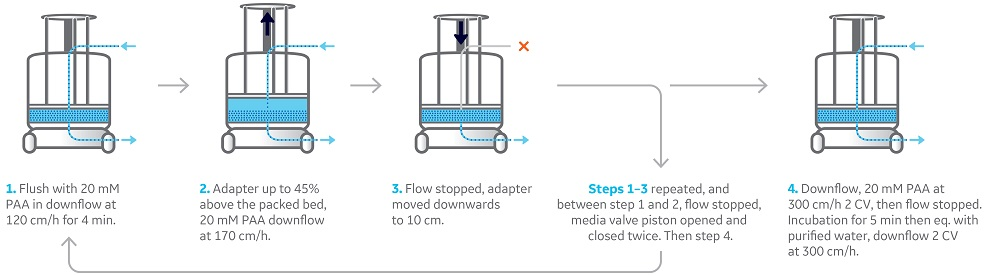
Fig 1. Graphical representation of sanitization method. Total time for method was 36 min for a 10.1 cm bed.
Sanitization of challenged column
The following sanitization method was run automatically by a programmed UNICORN method:
- Sanitization of a packed bed of 10.1 cm was initiated by a post-infection rinse of the packed bed with 2 CV of sterile purified water in a downflow at 60 cm/h.
- A rinse with 20 mM PAA in downflow at 120 cm/h for 4 min was performed, which forced the PAA solution into the packed bed.
- The adapter was moved upwards at 120 cm/h to 14.4 cm while 20 mM PAA was pushed through the bed downflow at 170 cm/h.
- Flow was stopped and the adapter moved downwards to 10.1 cm at 60 cm/h, pushing excess liquid out through the bottom mobile phase.
- A second rinse with 20 mM PAA in downflow at 120 cm/h for 4 min was performed, which forced the PAA solution further into the packed bed.
- The flow was again stopped, and the resin valve piston was open and closed twice within 6 s to clear the resin valve O-rings from trapped challenging organisms, from the contaminated resin filling.
- The adapter was moved upwards a second time at 120 cm/h to 14.4 cm while 20 mM PAA was pushed through the bed downflow at 170 cm/h.
- The flow was stopped, and adapter moved downwards a second time to 10.1 cm at 60 cm/h pushing excess liquid out through the bottom mobile phase. A rinse was performed by running a downflow with 20 mM PAA at 300 cm/h.
- The flow was then stopped and the PAA treated bed was incubated for 5 min to reach a resin contact time of ~ 35 min.
- Finally, the packed bed was equilibrated with purified water by running a downflow at 300 cm/h for 2 CV.
The automated sanitization steps are summarized earlier in Figure 1.
Liquid sample collection
Liquid samples were collected:
(1) after the consolidation phase of the packing before step 1;
(2) at a rinse with 2 CV of water after packing and before step 1;
(3) after equilibration with 2 CV of purified water in step 4.
After the sanitization method, the packed bed and resin valve were equilibrated with 20% ethanol and liquid samples were collected after 2 CV for the packed bed and 3 L for the resin valve. The 100 mM PAA in the rinse channel was rinsed out with ~ 150 mL 20% ethanol until pH increased and then a liquid sample was collected.
The column was then incubated with 20% ethanol for a 6-d clean hold while connected to the ÄKTAprocess system. Before any liquid samples were collected after the clean hold, the system was flushed with 20 mM PAA and equilibrated with 20% ethanol to minimize the risk of contaminants from the ÄKTAprocess system.
A liquid sample of the ethanol in the resin valve was collected and then 20% ethanol was run downflow at 60 cm/h through the packed bed and liquid samples were collected after 0.6 CV and 1.2 CV. A liquid sample was also collected from the top mobile phase of the filtered 20% ethanol as a control. The liquid in the flush channel was also collected.
Microbial sampling
After the clean hold, column disassembly was performed, and microbial samples were taken from the predetermined hardware sites as shown in Figure 2.
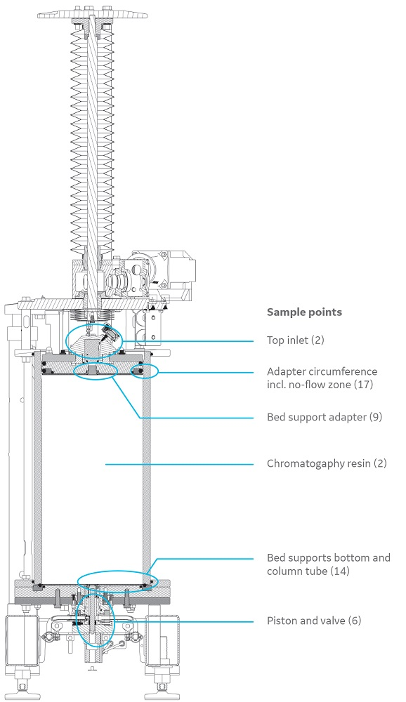
Fig 2. Predetermined liquid sampling points of the AxiChrom 300 column (numbers in brackets indicate the number of samples analyzed).
Microbial sampling was performed by one of the following methods:
1: air sampling
Sampling of air for airborne microorganisms to verify the surrounding hygiene level was conducted with a Microbial Air Sampler (MAS). The MAS loaded with an agar plate is positioned at a suitable measuring point. When measuring starts, a predefined volume of surrounding air is passed through the apparatus. Microorganisms are collected on the agar surface by impaction.
2: direct filtration
Sample solutions (minimum 10 mL) were collected in sterile tubes and then filtered through 0.45 μm cellulose nitrate membrane filters. Filters were incubated on agar plates at 30°C to 35°C for 5 d after which the plates were inspected for CFU.
3: swab
Surface samples were taken with swabs. The swab was inserted into the tube containing the isotonic swab rinse solution and vortexed for a minimum of 20 s. The solutions including the swabs were poured into Petri dishes and mixed with 30 mL of temperature controlled molten agar. Maximum temperature of the molten agar should be 45°C. After solidification, plates were incubated at 30°C to 35°C for 5 d after which the plates were inspected for CFU.
4: peptone water filtration
Detachable parts were aseptically removed and transferred to a sterile tube subsequently filled with 50 mL of sterile peptone water and then vigorously shaken for at least 20 min in room temperature (RT). The solutions were filtered through a 0.45 μm cellulose nitrate membrane filter. Filters were incubated on agar plates at 30°C to 35°C for 5 d after which the plates were inspected for CFU.
5: viable count
Samples of challenging organism suspensions were diluted in series in 0.9% NaCl. Samples from the diluted suspensions were plated on agar plates and incubated at 30°C to 35°C for 1 to 2 d after which the plates were inspected for CFU. The concentration of challenging organism was determined in the sampled suspensions.
6: agar plate
A sample of the chromatography resin was taken after sanitization and mixed with 30 mL of molten agar.
One gram of resin was aseptically transferred into a sterile container. The molten agar was aseptically added to the container and mixed with the resin to become homogenously suspended. Maximum temperature of the molten agar should be 45°C. The suspension was transferred to and allowed to solidify in Petri dishes. The plates were incubated at 30°C to 35°C for 5 d after which the plates were inspected for CFU.
Results from the sanitization procedure
Table 2 shows the results of aliquots collected before, during, and after packing as well as after the sanitization method.
Table 2. Microorganism levels (CFU/50 mL) at various stages of the sanitization study
| Sample | Flowthrough (FT) rinse/equil. with 2 CV of 20% ethanol | FT from resin valve bottom | Liquid from rinse channel equil. in 20% ethanol | Filtered 20% ethanol before application | FT 0.6 CV rinse/equil. with 20% ethanol | FT 1.2 CV rinse/equil. with 20% ethanol | FT from resin valve bottom (20% ethanol) | Liquid from rinse channel (20% ethanol) |
|---|---|---|---|---|---|---|---|---|
| Day 0 | 0 | 0 | 0 | - | - | - | - | - |
| Day 6 | - | - | - | 0 | 0 | 0 | 0 | 2* |
*Contaminants other than challenging organism, CFU/50 mL
Of the 68 samples analyzed in this study, no P. aeruginosa was found after sanitization (Table 2). Contamination with gram-positive Micrococcus luteus/lylae, which are airborne bacteria and common on the human skin, was observed in two samples, but at levels within the acceptance criteria of the study.
Before the sanitization study started, one specific part of the AxiChrom 300 design was thought to be more difficult to clean, that is, the no-flow zone formed between the top bed support and column tube. This design risk is not specific for AxiChrom 300 columns but may be a consequence of having a movable adapter. This risk is present in many column designs on the market.
The major difference with this sanitization study compared to previous studies in AxiChrom columns is that the homogenous resin slurry was first inoculated with the challenging organism and then packed in the column. This ensured that all critical parts of the column were challenged with a high concentration of P. aeruginosa. In previous studies the challenging organisms were applied through the mobile phase after the column had been packed.
The results here indicate that no contamination remained in the no-flow zone when the approach described was performed.
Because of the similarities in design of larger AxiChrom columns, it should also be possible to sanitize up to AxiChrom 2000 (2 m inner diameter) columns using this approach.
Bed performance after sanitization
The chromatography bed was evaluated for stability and performance after sanitization. To maintain bed flow properties as for a bed packed by the automated Intelligent Packing process for AxiChrom columns, bed compression by an additional 2 percentage points was required (Table 3). The bed performance was then equal to a bed packed by Intelligent Packing, although at somewhat higher compression. This was evaluated for two different bed heights and also in a larger AxiChrom 1000 column.
An alternative bed finalization before qualification is to fluidize the bed until completely homogenized and then pack the resin to its optimal compression, resulting in the same level of flow and bed performance at the target bed height as when packed directly by Intelligent Packing.
Table 3. Bed performance of 10 and 20 cm bed heights of MabSelect SuRe base matrix in AxiChrom 300 and 1000 before and after additional compression. Sanitization treatment corresponds to procedure under Sanitization of challenged column. Difference in flow properties is also shown comparing Intelligent Packing packed bed flow properties with bed subjected to the sanitization method and with additional bed compression
| Column | Theoretical plates (HETP/m) | Plate height, h (m) | Asymmetry factor, As | Max. flow velocity (cm/h) | |
|---|---|---|---|---|---|
| AxiChrom 300, 20 cm |
Initial performance before sanitization |
8200 |
1.4 |
1.1 |
650 |
| After sanitization |
6900 |
1.7 |
1.3 |
525 |
|
| AxiChrom 300, 20 cm |
Initial performance before sanitization |
7900 |
1.5 |
1.2 |
650 |
| After sanitization with PF1 increase of 2 percentage points |
5600 |
2.1 |
1.3 |
650 |
|
| AxiChrom 300, 10 cm |
Before sanitization |
7600 |
1.5 |
1.3 |
nd2 |
| PF increase of 2 percentage points; after sanitization |
6000 |
1.9 |
1.4 |
nd2 |
|
| AxiChrom 1000, 20 cm |
Initial performance, before sanitization |
8300 |
1.4 |
1.2 |
nd2 |
| After sanitization, with PF increase of 2 percentage points |
7600 |
1.5 |
1.4 |
nd2 |
2 nd = not determined
Conclusions
- 20 mM PAA (< 1 h) efficiently sanitized a bioprocess column packed with MabSelect SuRe base matrix.
- Number of challenging organism and other contaminants reduced from predetermined contamination levels to acceptable levels.
- Method duration reduced from 4 to 5 h using 1 M NaOH to 40 min with 20 and 100 mM PAA.
- Method automation ensures robustness and repeatability of sanitization.
- Finalizing the sanitized bed by either additional compression or fluidization and repacking gave identical performance to a bed packed using the Intelligent Packing method.
Trademark information
Ecolab is a trademark of Ecolab USA Inc.