Western blot membranes are critical to the success of your protein analysis workflow. Here, you’ll find tips on selecting the optimal hydrophobic Western blot membrane for your application.
Western blotting is a laboratory technique used to detect specific protein molecules in a protein mixture. You are able to evaluate the protein’s size, or estimate protein expression. Available since 1979, Western blotting remains an essential and fundamental analytical technique in many fields.
There are six steps involved in a general Western blotting protocol:
- Sample preparation
- Gel electrophoresis
- Proteins transfer
- Blocking
- Antibody incubation
- Protein detection and visualization
Most of these steps involve a microporous membrane that forms the solid support for your proteins. There are many different types of membranes that can be used in Western blotting protocols.
With various types of membranes available, it can be tricky to decide which is the best for your application. The two main membranes used for Western blots are nitrocellulose and polyvinylidene difluoride (PVDF). Both are hydrophobic, which allows proteins to bind to them.
In this blog, we compare nitrocellulose and PVDF. We go over their properties, and how each can affect your Western blotting results.
What do membranes look like?
Most people familiar with Western blotting will likely recognize nitrocellulose and PVDF membranes. If we take a closer look, there is an intricate 3D structure that confers their excellent capabilities as Western blot membranes.
Both nitrocellulose and PVDF membranes are classed as ‘tortuous path’ membranes. This means that they have pores with a random, interconnected sponge-like structure (Fig. 1). This structure can be manipulated in manufacturing to yield different pore sizes, making them suitable for various sizes of proteins.
While nitrocellulose and PVDF membranes may look similar, there are some significant differences, which affect their interaction with proteins and their application in Western Blot workflows (Table 1).
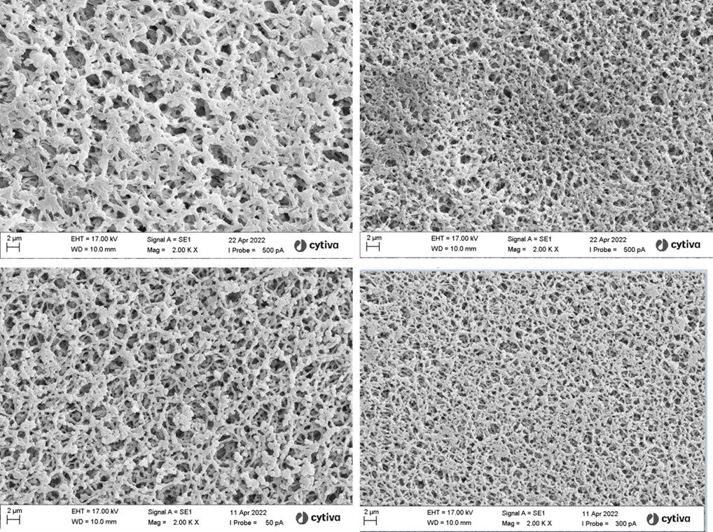
Fig 1. Electron micrographs of Western blotting membranes illustrating their 3D structure. (A) PVDF 0.2 μm, (B) PVDF 0.45 μm, (C) Nitrocellulose 0.2 μm, and (D) Nitrocellulose 0.45 μm.
How to choose the best membrane
The choice between nitrocellulose and PVDF membranes depends on your target protein(s), the selected detection strategy, and whether you intend to analyze multiple proteins through stripping and reprobing (Fig. 2).
Your target protein
Two properties to consider about your target protein are the abundance and size.
If your proteins aren’t particularly abundant, PVDF is the preferred choice because it has superior protein binding capacity and higher sensitivity. The sensitivity can be a drawback if you’re targeting high-abundance proteins as it can translate to higher background noise.
Nitrocellulose membranes are not capable of the detection sensitivity of their PVDF counterparts, but the lower background noise makes them ideal for proteins expressed at high levels.
Both membranes come in typical pore sizes of 0.1, 0.2, and 0.45 μm. While a 0.45 μm membrane is suitable for most protein blotting applications, smaller peptides or lower molecular weight proteins (< 15 kD) may require a 0.1 or 0.2 μm pore size membrane.
Table 1. Characteristics of nitrocellulose and PVDF membranes for Western blotting applications
| Nitrocellulose | PVDF | |
| Sample concentration | 80-100 µg of protein/cm2 | 150-200 µg of protein/cm2 |
| Protein size | Better for mid-to-low MW proteins | Better for high MW proteins |
| Protein binding interactions | Hydrophobic interactions | Hydrophobic and dipole interactions |
| Durability | Less durable | More durable |
| Saturation | Requires methanol in the transfer buffer | Requires methanol or ethanol prior to transfer |
| Chemical resistance | No, but improved by reinforced nitrocellulose membranes | Yes |
| Strip and re-probe | Possible, but can lose sensitivity during rounds. Reinforced nitrocellulose membranes improve suitability | Suitable |
| Autofluorescence | Low | High, but 'low-fluorescence' membranes are available |
| Detection | Well suited to chemiluminescence and fluorescence detection methods | Well suited to chemiluminescence detection but standard PVDF membranes can give high background. Dedicated low-fluorescence PVDF membranes can be used for fluorescence detection |
Your detection strategy
There are two common methods used in Western blotting experiments for detecting proteins bound to a membrane:
- Chemiluminescence: An indirect method for detecting proteins. Relies on an enzyme-substrate reaction that emits light.
- Fluorescence: Uses secondary antibodies directly conjugated to fluorescent dyes.
PVDF and nitrocellulose membranes are both compatible with chemiluminesnce-based protein detection methods. If you’re using fluorescence-based detection, a nitrocellulose membrane is needed due to the high autofluorescence of PVDF membranes.
Find out more about detection strategies for Western blots.
Stripping and re-probing
Stripping a Western blot is the method of removing primary and secondary antibodies from the membrane so it can be re-probed. In theory, a blot can be stripped and re-probed several times to visualize multiple proteins or to optimize methodology without needing to perform multiple gels and protein transfers.
Stripping the membrane involves harsh conditions that disrupts the interaction between the membrane-bound protein and the primary antibody. Nitrocellulose membranes are brittle and fragile so they can be difficult to strip and re-probe without losing signal. PVDF membranes offer a more durable and chemical-resistant material for multiple rounds of reprocessing.
Conflict resolution
Your needs for a Western blot membrane may be more complicated than the situations mentioned above.
Fortunately, some suppliers have developed membranes for these difficult circumstances.
If you have a low abundance protein and need to perform a fluorescence based Western blot, Cytiva™ offers dedicated “low-fluorescence” PVDF membranes, such as the Amersham™ Hybond™ LFP 0.2 µm PVDF membrane.
Nitrocellulose isn’t the first choice for stripping and re-probing, but is the better option for high-abundance proteins. Membranes such as the Amersham™ Protran™ 0.2 µm NC supported Western blotting membranes are made of reinforced nitrocellulose, which allows for multiple strip and re-probe cycles.
Figure 2 provides a key that can help guide your membrane selection for different Western Blotting applications.
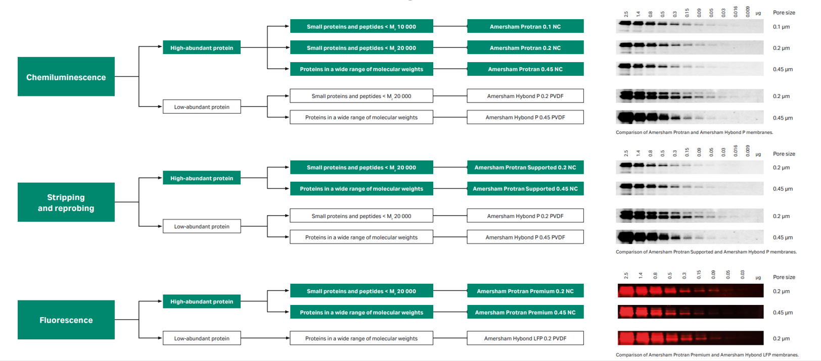
Fig 2. Western blotting membrane selection key.
Summary
Using the optimal membrane for your Western Blot application can be critical to your experiment’s success.
Learn more about how to select the best materials for your Western Blot.
Cytiva™ offers a range of Amersham™ Western blotting nitrocellulose and PVDF membranes, available in a variety of formats and pore sizes. If you have questions or would like to discuss your Western blotting workflow, contact our scientific support team.
Find the right Western blot membrane for your application here.