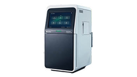
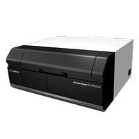
Gel and blot imaging is crucial to data interpretation. Blot imaging of Western blots allows for qualitative and quantitative evaluation of specific protein expression. By illuminating the membrane, identifying signals above background noise, capturing the image, and assessing relative intensities, Western blot imaging equipment allows the researcher to visualize the processed blots.
While classic Western blot processing on film required a darkroom and film development time, many current digital blot imaging systems can image the membrane immediately. Gel documentation systems, which show acrylamide gels after protein separation, are among the imagers used in Western blotting. Chemiluminescent imagers provide high sensitivity, signal stability, and ease of use.
After electrophoresis or immunodetection, you can use gel and blot imaging devices to view and examine gels and blots. These devices digitize signals, and the data is evaluated using sophisticated software before being saved for future use.